Peking University, April 30, 2018: Recently, Professor Chen Liangyi’s group in Institute of Molecular Medicine have developed an ultrasensitive super-resolution (SR) microscopy technique – Hessian structured illumination microscopy (Hessian SIM). The paper, titled as “Fast, long-term, super-resolution imaging with Hessian structured illumination microscopy”, was published as a full paper in Nature Biotechnology (https://www.nature.com/articles/nbt.4115).
At the temporal resolution of 188 Hz, Hessian-SIM is the fastest SR microscopy technology with 85 nm spatial resolution that can resolve 1/600 ~1/800 of one single hair. It is also the most photon efficient SR microscope that can operate at the photon dose 3 orders less than the classic point-scanning confocal microscope. Due to the ultralow photo-bleaching and photo- toxicity of Hessian SIM, we have achieved 97 Hz continous SR imaging in live cells for 10 min (equivalent to 180,000 SR images), or 1 Hz time-lapse SR imaging in live cells for one hour with mitigatable photo-bleaching.
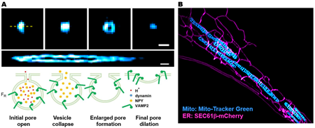
As compared to the stimulated emission depletion (STED) SR imaging technology that was awarded with 2014 Nobel Prize in Chemistry, Hessian SIM is superior in higher temporal resolution, ultra-low photo-toxicity that confers significant advantages in imaging live cell processes. For example, although both Hessian and STED (60 nm, 5 Hz, Shin W, et al., Cell. 2018) can resolve the enlarged fusion pore formed by the fusing vesicle with the plasma membrane, only Hessian SIM could detect four different fusion intermediates, including the initial opening of the fusion pore less than 3 nm, the collapsing of vesicle to the plasma membrane, the formation of enlarged pore, and the final pore dilation into the plasma membrane (Figure 1A).
Moreover, we are able to resolve mitochondrial cristae structures in live cells, and have visualized the structural dynamics of mitochondrial cristae during mitochondrial fusion and fission, and the internal re-organization of cristae structures within a single resting mitochondrion. With Hessian SIM, we are also able to visualize the dynamic interactions between mitochondria and endoplasmic reticulum in live cells (Figure 1B), which have never been observed by other SR imaging techniques such as STED and PALM/STORM. This again highlights the unique advantage of our ultrasensitive Hessian SIM in dynamically imaging photo-sensitive organelles such as mitochondria.
Figure 1: Hessian SIM enables novel structures and dynamics to be resolved in live cells. (A) The visualization of fusion intermediates resolved by Hessian SIM. We used VAMP2-pHluorin to label all insulin granules in INS-1 cells. (B) Dynamic interactions between mitochondria and endoplasm reticulum visualized by Hessian SIM. The COS-7 cell was transfected with SEC61b-mCherry, and labeled with Mito-Tracker Green dye.
The major technique breakthrough of Hessian SIM is a combination of hardware and algorithm innovations. On the hardware side, we have developed a novel segmented polarization rotator, precise control of the synchronization time sequences of the system as well as components that enable maximal emission detection. On the algorithm side, we propose for the first time, to use the continuity of biological structures in multiple dimensions as a priori knowledge to guide image reconstruction and attains artifact-minimized SR images. Overall, Hessian SIM is the fastest SR microscope and also sets the record in the length of continuous time-lapse SR imaging. Operating at a photo dose 1~3 order even less than point-scanning confocal microscopy, Hessian-SIM represents a big step for the SR microscopy to become a tool-of-chioce for biological researchers in daily experiments.
The first authors of the paper are Huang Xiaoshuai, Fan Junchao and Li Liuju, and the corresponding authors are Chen Liangyi and Tan Shan. The work was supported by grants from the National Science and Technology Major Project Program, National Natural Science Foundation of China, the Major State Basic Research Program of China, and Beijing Natural Science Foundation. Many authors of the project including Huang Xiaoshuai, Chen Liangyi, were also involved in the development of high spatiotemporal resolution miniature two-photon microscope that was published Nature Methods in 2017 and elected as “China's Top Ten Advances in Science in 2017”, “China's Top Ten Medical Science and Technology News in 2017” and “China's Top Ten Advances in Life Science in 2017”. In the future, they will also try to combine Hessian SIM with miniature two-photon microscopy for SR imaging in mice in vivo.
Source: Institute of Molecular Medicine
Edited by: Zhang Jiang