Peking University, Oct. 29, 2019: Polarization is a fundamental physical dimension of light, which has been exploited in the fields of light field modulation, quantum optics, stereoscopic filming, etc. In biology, the dipole orientation of fluorophores measured by polarization imaging reveals the orientation of targeted proteins. To study the protein orientation in subcellular structures, super-resolution polarization imaging has received increasing attention in recent years, while most techniques are still limited to labs with expertise.
Recently, Professor Xi Peng at Peking University College of Engineering and his colleagues developed polarized structured illumination microscopy (pSIM), which enables super-resolution polarization imaging on commercial SIM systems. This work was published in Nature Communications on October 16, 2019. (https://www.nature.com/articles/s41467-019-12681-w)
With pSIM, they extract the orientation information from off-the-shelf SIM systems with 2D-SIM, 3D-SIM, TIRF-SIM imaging capabilities. Compared with other polarization imaging methods, pSIM has the following three advantages at the same time: high spatial resolution for ultrastructure; resolving dipole orientation of biomolecules; fast dynamics imaging in live cells.
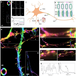
Figure 1 PSIM reveals a new model of actin ring "side by side" assembly in MPS, which overturns the textbooks hypothesis of "end-to-end" structure.
Biologists go for structured illumination microscopy for its high resolution and fast imaging speed. In most SIM systems, two linearly polarized light interferences to generate structured illumination. The polarization modulation of the interfering light in SIM needs to be high for better reconstruction, which making SIM itself a natural polarization imaging system. The researchers delivery many biological experiments to demonstrate its broad applicability, such as λ-DNA, the actin filaments in BAPE cells and mouse kidney tissue, the interaction between actin and myosin, and GFP-stained microtubules in live U2OS cells.
Furthermore, the team studied the membrane-associated periodic skeleton (MPS) in neurons. pSIM overturned the textbook “end-to-end” actin ring structure hypothesis with high spatial resolution and accurate polarization detection, and revealed the new “side-by-side” assembly model of actin ring in MPS . With the high spatial-temporal resolution inherited from SIM and the unique dipole orientation information, pSIM is highly promising in the application of answering a wide variety of biological questions in the future.
An innovative technology usually takes the following two approaches to benefit the scientific research community: 1) open access, so that other scholars can get access by building similar systems; 2) commercialize, so that others can get access by purchasing instruments. This work has opened up the third approach to promote scientific research: through in-depth re-examination of the potential characteristics of SIM technology, they discovered the polarization detection characteristics of the existing SIM systems, an important character that even the inventor did not realize, so that the existing systems can be upgraded to polarization SIM system instantly, without any change in hardware. This enables the life science laboratories with SIM system to directly access polarization SIM, which can greatly promote the research of polarization super-resolution imaging.
In recent years, the research group of Xi Peng has been committed to the development of polarization super-resolution technology and SIM super-resolution technology, such as: 1) Super-resolution dipole orientation mapping (SDOM) was published in the Journal of light: Science and applications, and was highlighted by Nature methods (http://news.pku.edu.cn/xwzh/129-295505.htm); 2) SDOM was applied to SERS super-resolution imaging of gold nanoparticles (Nanoscale 2018); 3) reduced frame SIM to improve the speed of structured light imaging by over two times (IEEE tip2018); 4) participated in the development of Hessian SIM ultra-high speed structured light imaging technology, and proposed rolling SIM, which can improve the imaging speed of SIM by more than three times (Nature Biotechnology 2018). These work have laid a solid foundation for pSIM.
The co-corresponding authors of this work are Dr. Xi Peng of Peking University and Dr. Dai Qionghai, a professor of Tsinghua University and member of Chinese Academy of Engineering. Dr. Karl Zhanghao, the co-first author and co-corresponding author of this work, is supported by the Boya Post doctoral program of Peking University. Chen Xingye, the co-first author, is a doctoral student of Automation Department of Tsinghua University. The neural cell experiment was completed by Professor Zhang Yan of McGovern Brain Science Center, Peking University. The in vitro actin experiment was completed by Professor Li Xiangdong of Institute of Zoology, Chinese Academy of Sciences. The living cell microtubule imaging was completed by Professor Chen Xiaowei, School of Life Sciences, Peking University. The SIM super-resolution micro imaging of this work was completed on the biological microscopy platform of Peking University, with the help of Dr. Shan Chunyan and others. This work is supported by the National Natural Science Foundation of China, the Ministry of science and technology, the National Science Foundation for of Beijing Municipal Science and Technology Commission under Beijing Distinguished Young Scholars Program, the Med + X Project and the Instrumentation Project of Peking University.
Edited by: Huang Weijian
Source: College of Engineering, Peking University