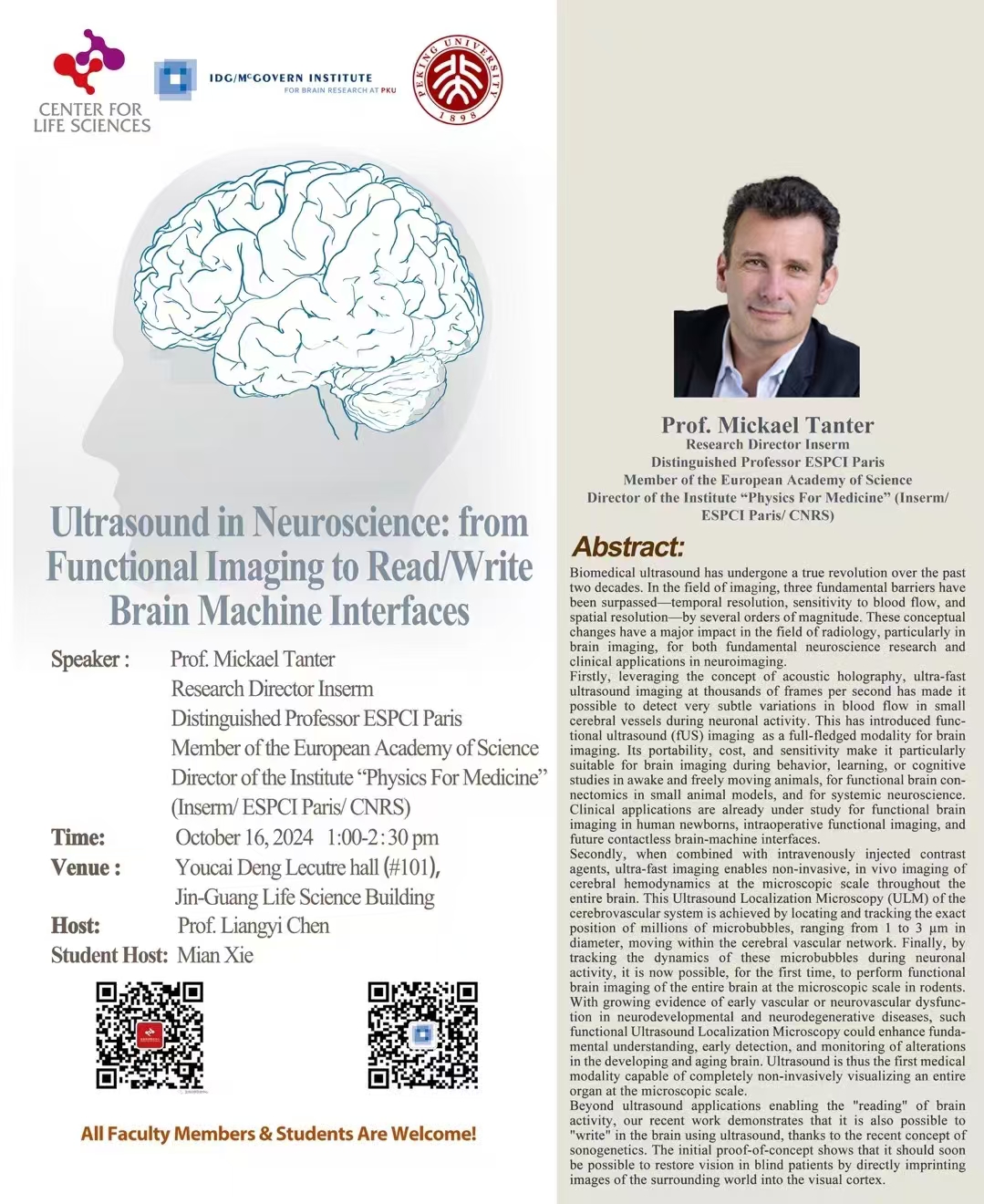
Speaker: Prof. Mickael Tanter Research Director Inserm Distinguished Professor ESPCI Paris Member of the European Academy of Science Director of the Institute "Physics For Medicine" (Inserm/ ESPCI Paris/ CNRS)
Time: 13:00 - 14:30 p.m., Oct 16, 2024, GMT+8
Venue: Youcai Deng Lecutre hall (#101), Jin-Guang Life Science Building, PKU
Abstract: Biomedical ultrasound has undergone a true revolution over the past two decades. In the field of imaging, three fundamental barriers have been surpassed temporal resolution, sensitivity to blood flow, and spatial resolution- by several orders of magnitude. These conceptual changes have a major impact in the field of radiology, particularly in brain imaging, for both fundamental neuroscience research and clinical applications in neuroimaging.
Firstly, leveraging the concept of acoustic holography, ultra-fast ultrasound imaging at thousands of frames per second has made it possible to detect very subtle variations in blood flow in small cerebral vessels during neuronal activity. This has introduced functional ultrasound (fUS) imaging as a full-fledged modality for brain imaging. Its portability, cost, and sensitivity make it particularly suitable for brain imaging during behavior, learning, or cognitive studies in awake and freely moving animals, for functional brain con-nectomics in small animal models, and for systemic neuroscience.
Clinical applications are already under study for functional brain imaging in human newborns, intraoperative functional imaging, and future contactless brain-machine interfaces.
Secondly, when combined with intravenously injected contrast agents, ultra-fast imaging enables non-invasive, in vivo imaging of cerebral hemodynamics at the microscopic scale throughout the entire brain. This Ultrasound Localization Microscopy (ULM) of the cerebrovascular system is achieved by locating and tracking the exact position of millions of microbubbles, ranging from 1 to 3 um in diameter, moving within the cerebral vascular network. Finally, by tracking the dynamics of these microbubbles during neuronal activity, it is now possible, for the first time, to perform functional brain imaging of the entire brain at the microscopic scale in rodents. With growing evidence of early vascular or neurovascular dysfunction in neurodevelopmental and neurodegenerative diseases, such functional Ultrasound Localization Microscopy could enhance fundamental understanding, early detection, and monitoring of alterations in the developing and aging brain. Ultrasound is thus the first medical modality capable of completely non-invasively visualizing an entire organ at the microscopic scale.
Beyond ultrasound applications enabling the "reading" of brain activity, our recent work demonstrates that it is also possible to "write" in the brain using ultrasound, thanks to the recent concept of sonogenetics. The initial proof-of-concept shows that it should soon be possible to restore vision in blind patients by directly imprinting images of the surrounding world into the visual cortex.
Source: McGovern Institute for Brain Research at PKU
